Toxins
In Skin Bacteria |
From
Alan Cantwell MD |
| Toxins In Skin Bacteria Help Lymphoma Cells Spread
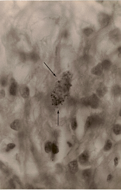
Figure 1. Hodgkin’s (B cell) lymphoma of the lung. Arrows point to a rare collection of intracellular variably staining coccoid forms. MacCallum-Goodpasture Gram stain, magnification x1000, in oil.
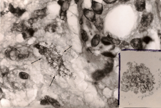
Figure 2. Hodgkin’s lymphoma of the skin. Arrows point to a large collection of minute granules and coccoid forms in the deep dermis. Fite (acid-fast) stain, x1000, in oil. Insert shows appearance of Propionibacterium (Corynebacterium) acnes bacterial cultured from the skin lesion. Ziehl-Neelsen (acid-fast) stain, x1000, in oil Compare the similar size and shape of the bacteria seen in the skin (in vivo) with those cultured in the laboratory.
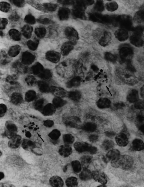
Figure 3. Hodgkin’s lymphoma of the lymph node. Variably sized coccoid forms and larger bodies consistent with cell wall deficient “large bodies” and also to “Russell bodies.” Gram stain, x1000, in oil.
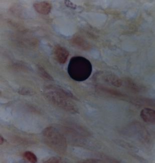
Figure 4. Hodgkin’s lymphoma of the lymph node showing very large solitary “large body” (similar to a Russell body). Gram stain, x1000, in oil.
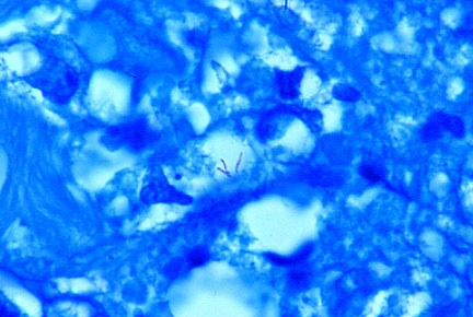
Figure 5. Immunoblastic sarcoma tumor (renamed immunoblastic lymphoma) of the skin in an advanced case of AIDS. Two very rare acid-fast (red stained) rod-like bacillary forms are seen in the center. Fite stain, x1000, in oil.
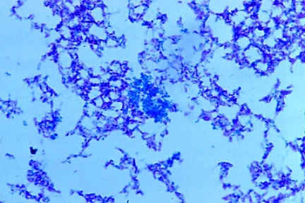
Figure 6. Mycobacterium avium intracellulare cultured from the immunoblastic sarcoma/lymphoma facial tumor. Note the pleomorphic forms, such as acid-fast rods and non-acid-fast (blue-stained) cocci. Ziehl-Neelsen (acid-fast) stain, x1000, in oil.
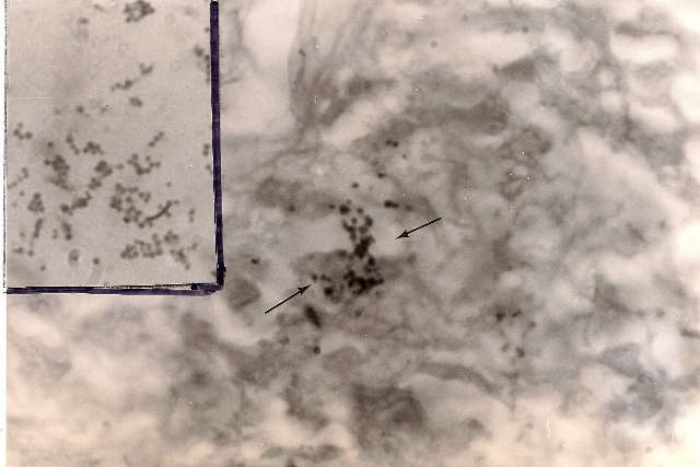
Figure 7. Mycosis fungoides (cutaneous T-cell lymphoma) of the skin. Arrows point to extracellular scattered coccoid forms in the deep dermis. Fite-Faraco (acid-fast) stain, x1000, in oil. Insert shows Staphylococcus epidermidis cultured from the lesion. Note the similar size and shape of the cocci cultured to the coccoid forms seen in vivo in the skin lymphoma. Ziehl-Neelsen (acid-fast) stain, x1000, in oil.
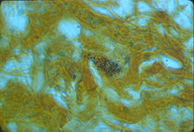
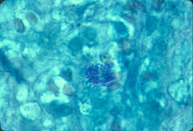
Figure 8. Mycosis fungoides (T-cell lymphoma) of the skin showing a large clump of closely-knit coccoid forms in the dermis. Alexander-Jackson’s intensified triple stain for the detection of non-acid-fast forms of mycobacteria, x1000, in oil.
Figure 9. Mycosis fungoides (T cell lymphoma) of the lymph node showing an intracellular clump of tightly-packed coccoid forms. Alexander-Jackson’s triple stain, x 1000, in oil. Compare the size and shape of the node forms to those seen in the skin in Figure 7 from the same patient. |
| Donate to Rense.com Support Free And Honest Journalism At Rense.com | Subscribe To RenseRadio! Enormous Online Archives, MP3s, Streaming Audio Files, Highest Quality Live Programs |
