Look at the incredibly intricate components within a single nanotech fiber
like are now being found in PCR test kit swabs and face masks.
These photos of a fiber under 5600x magnification were taken in 2006.
Imagine where the nanotechnology is now...
Magnification approximately 5600x.
Notice internal filament structure within the fiber.
Width of the internal fibrous structure is at the micron or sub-micron level.
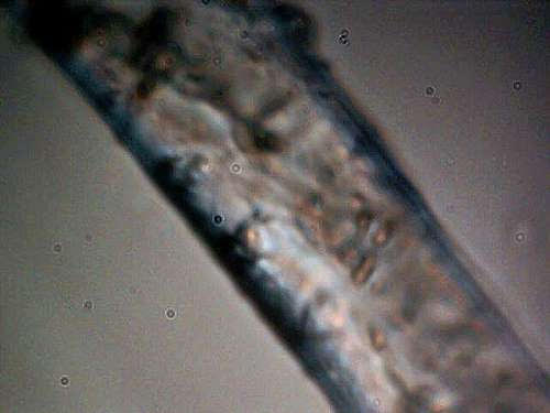
Magnification approximately 5600x.
Notice internal generally circular structures.
Strongly indicative of a biological nature at this point.
These structures measure on the order of 1 micron (virus size).
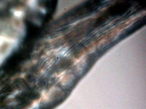
Increasingly complex internal nature of the original sample fiber is now evident.
Magnification approximately 5600x.
Complex internal organization of sub-fibers and structural forms is apparent.
Magnification approximately 5600x.
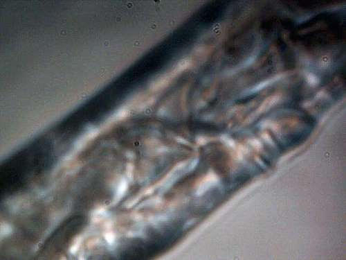
This photograph shows the ability of the fiber to be folded and/or twisted.
Internal parallel organization of sub-fibers is visible.
Non-uniformity of the fibers dimensions is also evident.
Transverse separation or structure also visible in lower right of image.
Magnification approximately 5600x.
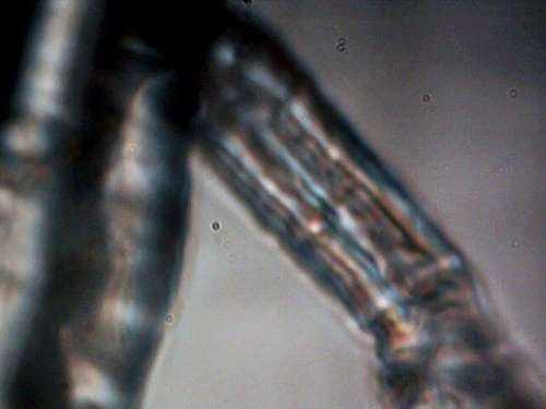
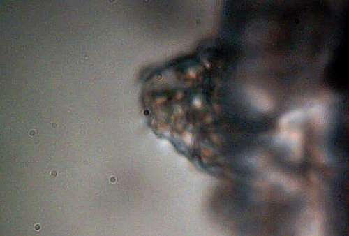
Additional budding structure visible on the edge of the primary fiber.
Complex internal micron size structures within.
Translucent encasement that is indicative or suggestive of reproductive capability.
Magnification approximately 5600x.
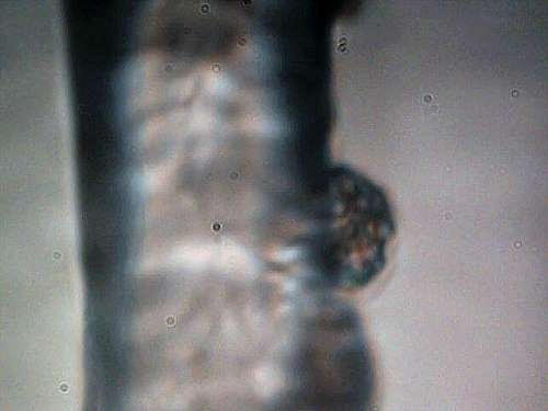
Additional budding structure visible on the edge of the primary fiber.
Complex internal micron size structures within.
First © 2006 by Jan Smith and Clifford E Carnicom