|
The
cause of breast cancer is unknown, but over the past century various
controversial scientists have implicated bacteria as the causative agent
[1-8]. Despite this cancer microbe research, the bacterial theory of
cancer has been widely rejected. This report suggests that the elimination
of bacteria as breast cancer-causing agents was premature; and that
new research gives additional credence to bacteria causing breast cancer.
The
findings of the Human Microbiome Project, launched in 2006,
indicate the human body contains 100 trillion microbes, most of
which are bacteria. In fact, 90% of the cells of the body are not
human cells, but microbial cells. The precise role these bacteria
play in cancer is not known, nor has it been studied.
In
addition, only recently have we learned that various organs and
tissue of the human body are not
sterile. Previously it was believed that such tissue was devoid of
bacteria. Now various bacteria have been detected by molecular
biology techniques and properly stained microscopic examination. In
2014 Urbaniak et al. [9] found a diverse population of bacteria in
breast tissue, the details of which can be found online at:
http://www.ncbi.nlm.nih.gov/pmc/articles/PMC4018903/
There
are many different risk factors postulated for breast cancer, but
according to the National Cancer Institute, breast cancer is a
genetic disease caused by certain changes to genes that control the
way our cells function, especially how they grow and divide. These
changes include mutations in the DNA that make up our genes. In this
regard, it is well-accepted that bacteria frequently swap genes
with one another. More controversial is recent research indicating
that bacteria can also exchange genes with human cells, especially
cancer cells. For this reason, the presence of bacteria in cancer
should be taken seriously as a source of genetic alteration and
cancer production [10].
Bacteria found in cancerous
tissue have generally been dismissed as secondary invaders of tissue
weakened by cancer. Bacteria cultured from cancerous tissue are often
interpreted as laboratory contaminants of no etiologic consequence.
With the new knowledge that various bacteria are universally present
in breast tissue, their presence in cancer can no longer be assumed
to be secondary. In fact, bacteria are always present before
the cancer process is initiated. The reported microbiology of cancer
indicates that cancer bacteria often grow in the lab as common bacteria,
such as staphylococcal, streptococcal, and corynebacteria-like bacteria.
Because bacteria are generally thought to play no role in cancer, such
isolates are generally regarded as contaminants having no significance.
According to
cancer microbe researchers, the germ is a pleomorphic,
intracellular and extracellular microorganism that can be detected in
cancerous tissue by the use of special tissue stains, particularly
the acid-fast stain traditionally used for the detection of TB
mycobacteria. The common form in tissue is the round
staphylococcus-like coccoid form. These coccoid forms vary in size
from barely visible granules up to much larger globoid
forms [1,2,4,7].
The following
5 photos show the appearance of these coccoid forms in a case of intraductal
breast cancer previously reported in 1981 [8].
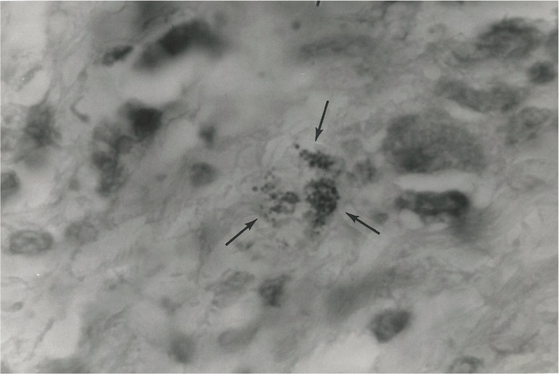
Figure
1. Tissue section of breast cancer showing tumor cells and a nest of
extracellular coccoid forms and still smaller granular forms, some
of which are barely visible. Intensified Kinyoun (acid-fast) stain,
magnification x 1000, in oil.
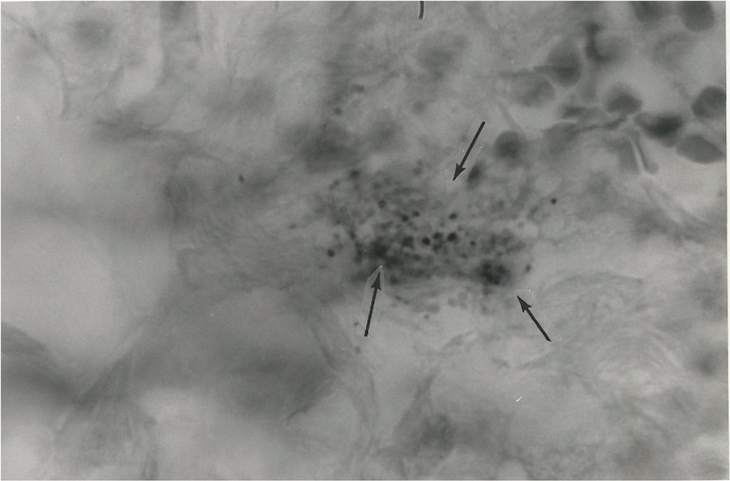
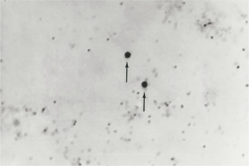
Figure
2. Tissue section of breast cancer showing a nest of extracellular variably-sized
coccoid forms. A collection of red blood cells is seen in the upper
right. Intensified Kinyoun stain, x 1000, in oil.
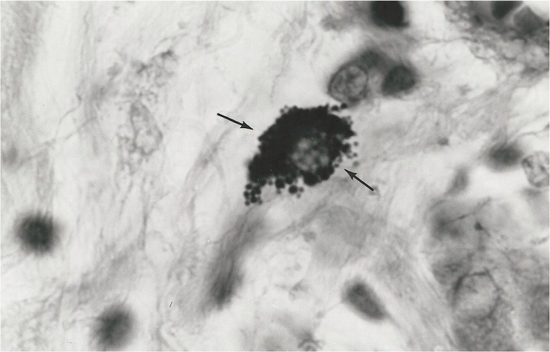
Figure
3. Tissue section of breast cancer showing intracellular variably-sized
coccoid forms and larger globoid forms. Intensified Kinyoun stain. x
1000, in oil
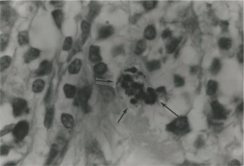
Figure
4. Tissue section of breast cancer showing a nest of variably-sized
coccoid forms ranging in size from barely visible "granules"
up to the size of ordinary staphylococci and still larger "globoid"
forms. The size range and pleomorphic nature of the round forms suggest
growth forms of so-called cell wall deficient bacteria. Intensified
Kinyoun stain, mag x 1000, in oil.
Figure
5. Smear of Staphylococcus
epidermidis
cultured from a biopsy taken from a metastatic nodule when the original
breast cancer tumor metastasized to the skin. All the coccal forms in
this photo are the size of ordinary cocci, except for two much larger
forms consistent with the size of globoid forms. Compare the size and
shape of the coccoid forms found in the breast tumor in photos 1-4 with
the size and shape of the staphylococci grown in the laboratory. Gram
stain, x 1000, in oil.
Why have bacteria in breast tissue and breast
cancer been ignored or overlooked for a century? One explanation is
that the pathologists routine tissue staining methods for
cancer diagnosis does not demonstrate bacteria well. Cancer microbe
researchers discovered that special stains were required,
particularly the acid-fast stain [1,4,7]. Also the cancer microbe is
pleomorphic and can exhibit various guises in tissue and in the
laboratory [7]. Such bacteria have been described as cell wall
deficient bacteria, and there is also an ultramicroscopic viral-size
growth form that can pass through lab filters designed to hold back
bacteria [4].
For
more details on cancer microbe research, see my articles on
rense.com, including Bacteria cause cancer; The microscopic
evidence and Cancer is an infection caused by tuberculosis-type
bacteria at http://www.rense.com/general95/bacmicro.html
and http://www.rense.com/general80/canc.htm
Currently,
there is a slight renewal of interest in bacteria and cancer. In an
editorial entitled Cancer and the microbiome, published in
April 2015 in Science,
Dr. Wendy Garrett explains how microbes could influence
carcinogenesis by causing changes in cell proliferation and death;
through immune system interference; and via metabolism of food,
pharmaceuticals, and host-produced chemicals [11].
At
present, the serious research of cancer microbe workers that took
place decades ago has been largely forgotten, and rarely, if ever,
cited by current researchers of the microbiome. This is a tragic (as
well as unscientific) situation.
Bacteria are intimately associated with inflammation
in the body; and inflammation precedes cancer. Bacteria are always present
in breast tissue. Thus, their presence always precedes the growth of
cancer. Bacteria should surely be given some renewed consideration as
a cause of breast cancer.
SELECTED
REFERENCES
1.
Wuerthele Caspe (Livingston) V, Alexander-Jackson E, Anderson JA, et
al: Cultural properties and pathogenicity of certain microorganisms
obtained from various proliferative and neoplastic diseases. Amer
J Med Sci 220:628-646,
1950.
2.
Alexander-Jackson
E: A specific type of microorganism isolated from animal and human
cancer: Bacteriology of the organism.Growth 18:37-51,
1954.
3.
Diller IC: Growth and morphologic variability of pleomorphic,
intermittently acid-fast organisms isolated from mouse, rat, and
human malignant tissues. Growth 26:181-209,
1962.
4.
Wuerthele-Caspe Livingston V, Alexander-Jackson E: An experimental
biologic approach to the treatment of neoplastic disease.J
Amer Med Women's Assn 20:858-866,
1965.
5.
Seibert FB, Farrelly FK, Shepherd CC: DMSO and other combatants
against bacteria isolated from leukemia and cancer patients. Ann
NY Acad Sci 141:175-201,
1967.
6.
Seibert FB, Yeomans F, Baker JA, et al: Bacteria in tumors. Trans
NY Acad Sci 34(6):504-533,
1972.
7.
Wuerthele Caspe Livingston V, Livingston AM: Some cultural,
immunological, and biochemical properties of Progenitor
cryptocides. Trans
NY Acad Sci 36(6):569-582,
1974.
8.
Cantwell AR Jr, Kelso DW: Microbial findings in cancer of the breast
and in their metastases to the skin. J
Dermatol Surg Oncol 7:483-491,
1981.
9.
Urbaniak C, Cummins J, Brackstone M, et al. Microbiota of human
breast tissue. Appl
Environ Microbiol
(2014) May;80(10):3007-14. doi: 10.1128/AEM.00242-14. Epub 2014 Mar
7.
10.
Riley DR, Sieber KB, Robinson KM, et al. Bacteria-Human Somatic Cell
Lateral Gene Transfer Is Enriched in Cancer Samples. Eisen JA, ed.
PLoS
Computational Biology.
2013;9(6):e1003107. doi:10.1371/journal.pcbi.1003107.
11.
Garrett WS. Microbial dysbiosis is associated with breast cancer. PLoS
One
(2014); 9(1): e83744. Published online 2014 Jan 8. doi: 10.1371/journal.pone.0083744.
[Alan
Cantwell is a retired dermatologist. He is the author of The
Cancer Microbe and Four Women Against Cancer, available from amazon.com
Email:
AlanRCan@aol.com]
|